Mechanical Models for 50-Year Lifetime PV Modules
DuraMAT focuses on the development of material behavior models that can be used to address long-term degradation issues in photovoltaic (PV) modules.
The viscoelastic behavior of various polymeric components will be characterized and fit to existing models (i.e., Generalized Maxwell model), and their respective Prony Series will be made publicly available for direct input into finite-element analysis software. Additionally, the long-term, wear-out mechanisms of solar cell gridlines will be examined.
Through scanning electron microscopy with in-situ mechanical loading, the driving forces behind wear-out fatigue of gridlines will be identified, and a subsequent probabilistic failure model will be developed. The model will be experimentally validated using the dynamic mechanical loading tool developed in previous DuraMAT efforts.
Core Objective
Multi-Scale, Multi-Physics Model
Location
National Renewable Energy Laboratory
Applications
The material behavior models for laminate materials could be applied to finite-element analysis problems to address a number of thermomechanical-related issues and scenarios for PV modules. Applications might include the study of hail-impact or interconnect fatigue.
Availability
This capability is available to all DuraMAT collaborators.
References
N. Bosco, M. Springer, and X. He, "Viscoelastic Material Characterization and Modeling of Photovoltaic Module Packaging Materials for Direct Finite-Element Method Input," IEEE Journal of Photovoltaics, vol. 10, no. 5, pp. 1424-1440, 2020
T. Silverman, M. Bliss, A. Abbas, T. Betts, J. Walls, and I. Repins, Movement of Cracked Silicon Solar Cells During Module Temperature Changes, 2019 46th IEEE Photovoltaic Specialists Conference
N. Bosco, A. Chavez, V. Upadhyaya, and S. Han, Fatigue-Like Behavior of Silver Metallization Gridlines and Proposed Damage Mechanics Model, 2020 47th IEEE Photovoltaic Specialists Conference
Contact
To learn more about this capability, contact Michael Owen-Bellini or Nick Bosco.
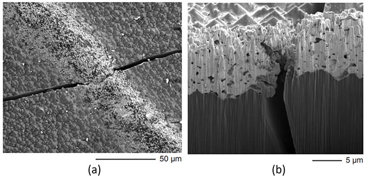
Scanning electron microscopy images from (a) the top and (b) at the cross-section of a cell crack, showing the silver metallization bridging the cell gap.